DSS: Redefining Biotechnology & Life Science in India


Home > Products & Services > Software > PathFusion
PathFusion
FISH | Tissue Matching | Whole Slide Imaging
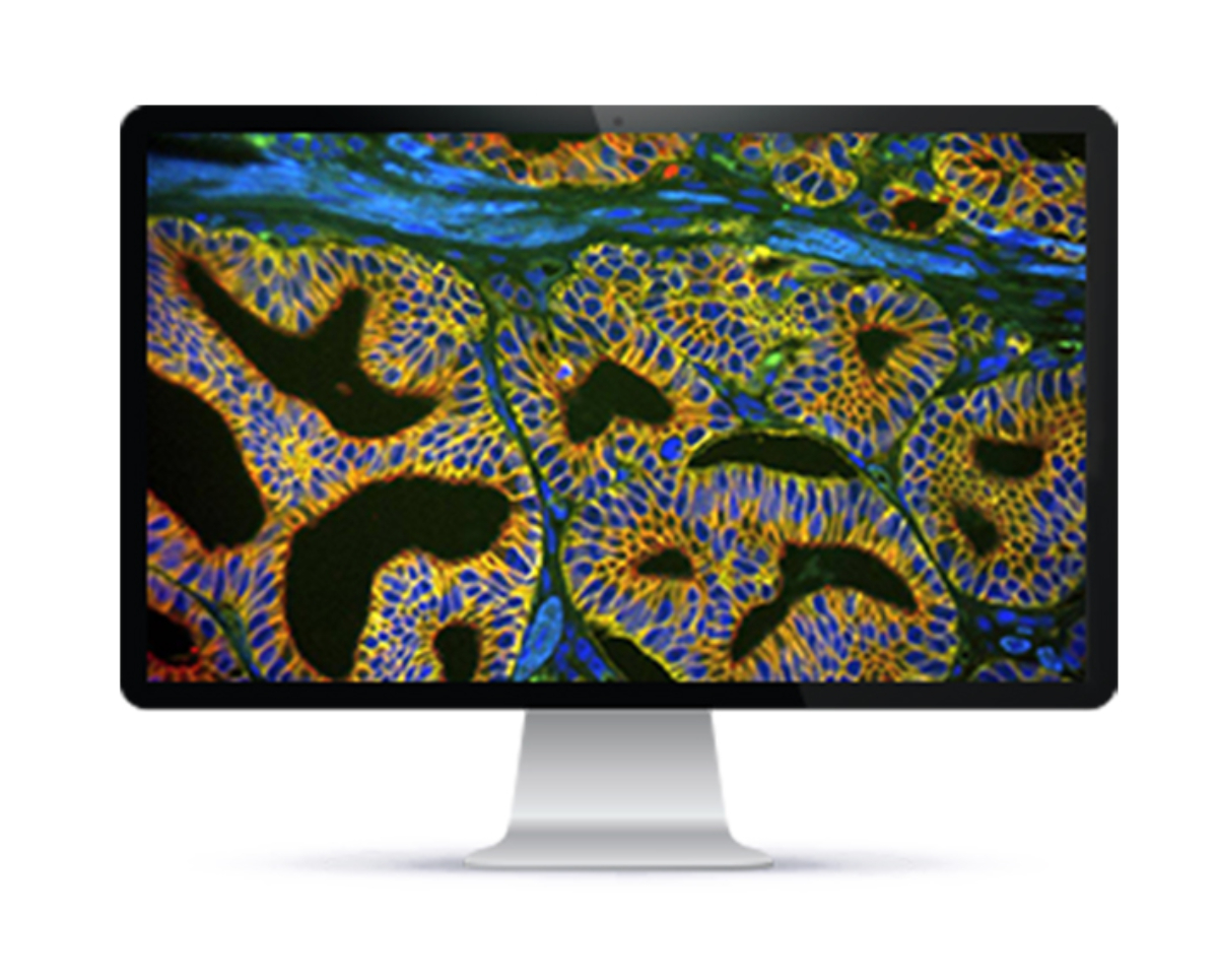
PathFusion is a state-of-the-art diagnostics solution that combines several advanced imaging technologies to provide highly accurate and validated analysis for better diagnostic confidence. The suite includes Fluorescence In Situ Hybridization (FISH), Tissue Matching, and Whole Slide Imaging (WSI), which can be used together or separately depending on the diagnostic needs.
FISH is a technique used to detect and locate the presence or absence of specific DNA sequences in cells or tissues. It involves the use of fluorescent probes that bind to specific DNA sequences and emit light when exposed to a specific wavelength of light. FISH is a highly sensitive and specific technique that can detect genetic aberrations in cancer cells that are not visible using traditional staining methods.
Tissue Matching is a revolutionary digital imaging technology that allows the matching of FISH results with H&E/IHC-stained tissue samples. This allows for a more accurate and comprehensive analysis of genetic aberrations in cancer cells, which can help to guide treatment decisions and improve patient outcomes.
WSI is a digital imaging technique that allows the capture of high-resolution images of entire tissue sections. These images can be viewed and analyzed on a computer screen, allowing for detailed examination of tissue morphology and cellular structures. WSI is a powerful tool for pathology diagnosis, as it allows for remote consultation and collaboration between pathologists, and can also be used for educational and training purposes.
Overall, the combination of FISH, Tissue Matching, and WSI in the PathFusion suite offers a comprehensive and cutting-edge diagnostic solution for cancer patients, providing accurate and validated analysis for higher diagnostic confidence.
About
ASI Imaging Instruments in India
Applied Spectral Imaging develops computer-aided systems for use in diagnostics by pathology, cytogenetic and research laboratories and helps provide labs with accurate, repeatable and standardized analysis of karyotyping, FISH, CISH, quantitative IHC, as well as SKY (spectral karyotyping) spectral imaging for research applications. Their further presence in India is led by DSS Imagetech.
More Products
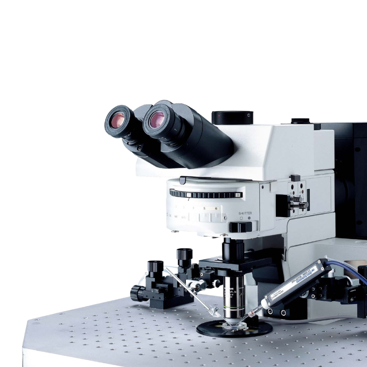
BX51WI
The Olympus BX51WI is a high-performance upright microscope meticulously designed for advanced in vivo and…
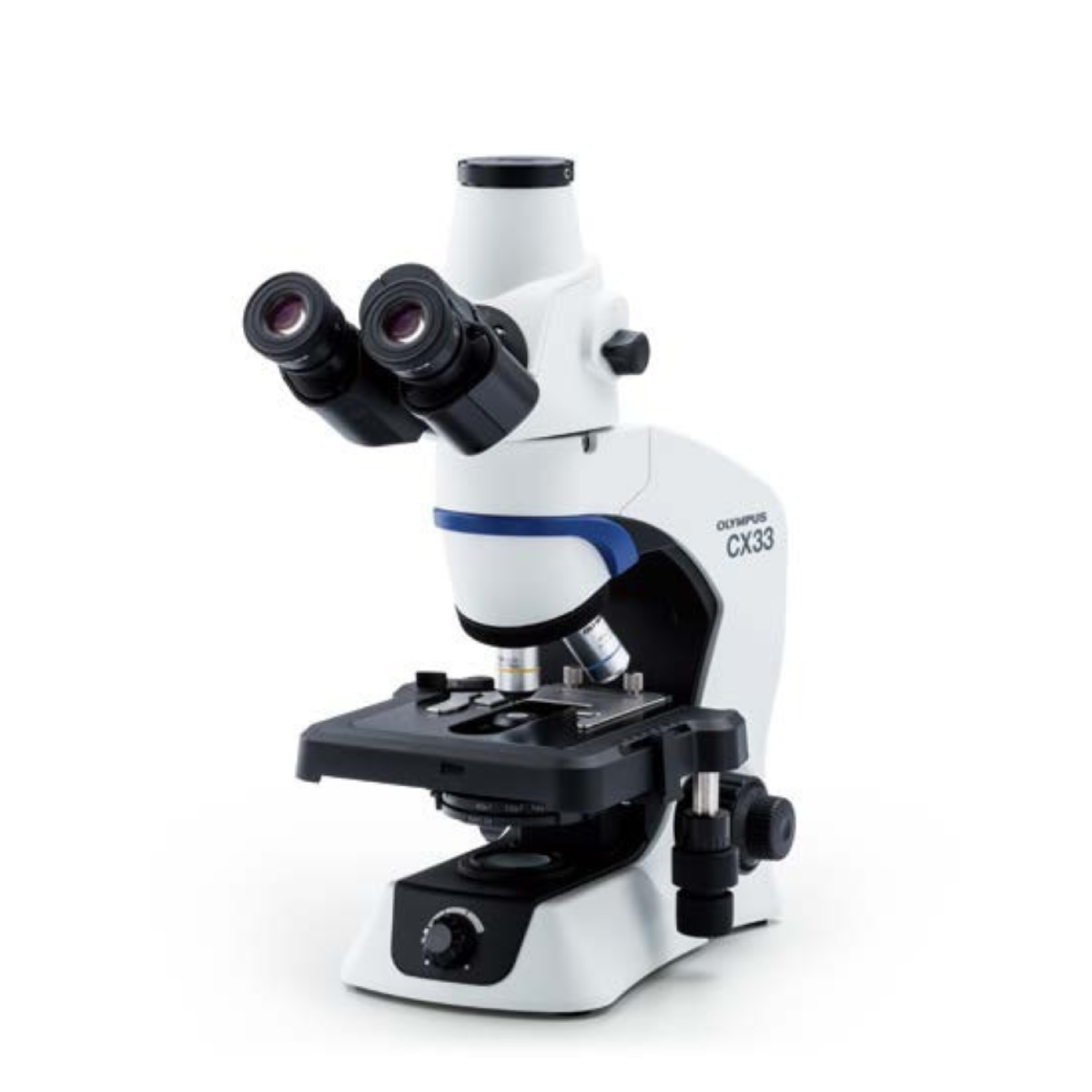
CX33
Designed to streamline everyday laboratory routines, the Olympus CX33 delivers clear, bright images with minimal…
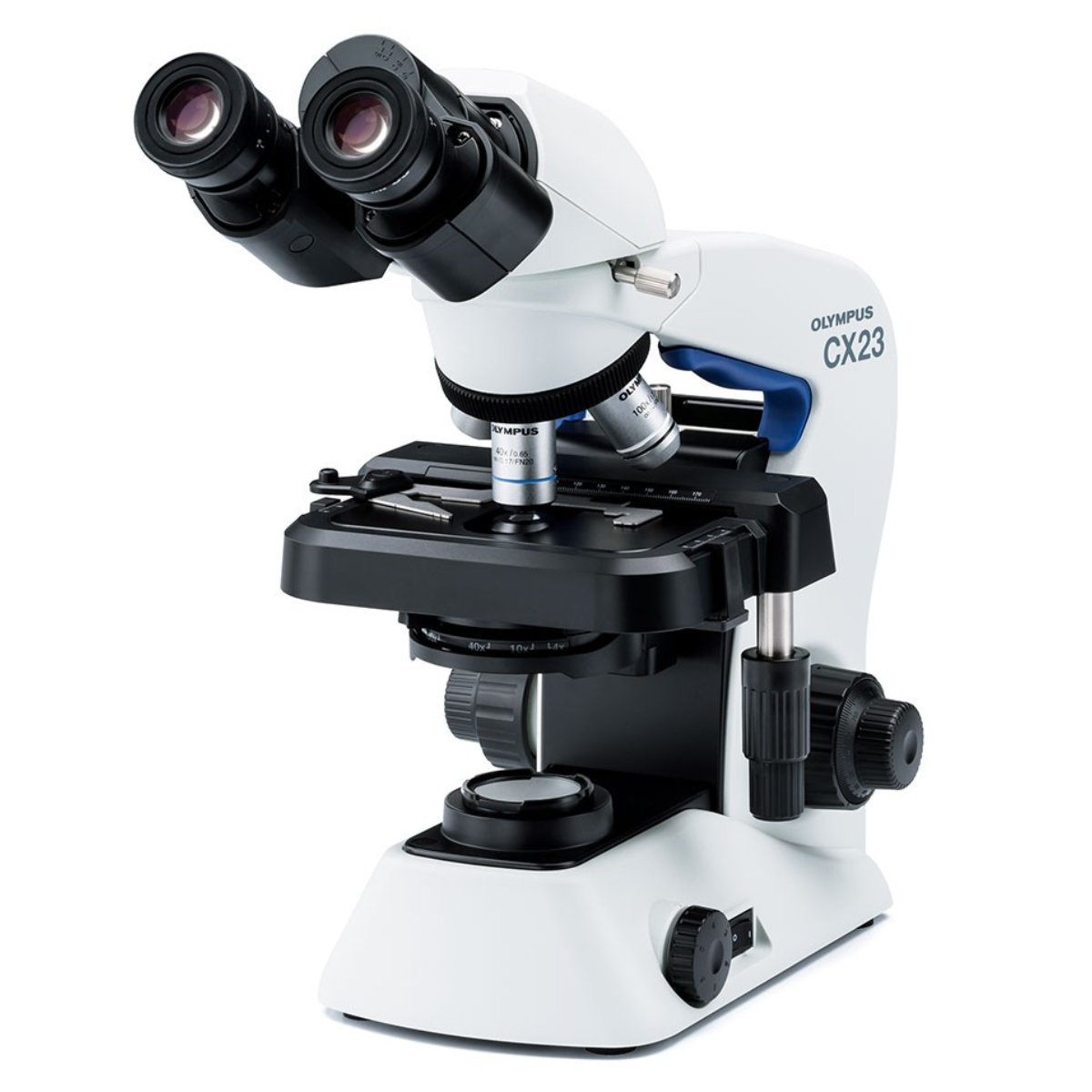
CX23
Designed for operational ease, the CX23 microscope’s unique features accommodates the student and every requirement…
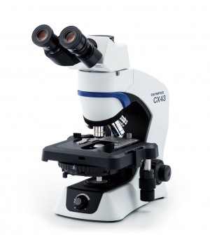
CX43
The CX43 microscope enable users to remain comfortable during long periods of routine microscopy observations.…
Testimonials & Reviews
Dr. Chhaya Chande, Professor & HOD, Microbiology
GGMCJJ Hospitals, Mumbai
“Ms. Megha Dhumal (Assistant Manager- Application) has done a satisfactory demonstration of the running of the Abbott Sample preparation machine model m2000sp and the Abbott RT-PCR machine model m2000rt. We appreciate the effort made by the DSS team under these difficult conditions to help our lab to carry out the imperative Covid-19 tests.”
Dr Sunil K Arora, Professor, Deptt of Immunopathology
PGIMER, Chandigarh
“We are using Confocal Microscope and one Fluorescence Microscope. Both are working fine. The after sales services by DSS have been excellent for functioning & upkeep of the microscopes. The applications support by experts from DSS is very useful. Keep it up!”
Dr Pramod Kumar Bajaj
MD, Spermprocessor Pvt Ltd
“Really excited to see the DSS Pathology solutions exhibition booth at APCON 2019 along with Magnus. We think all the upcoming technology had been displayed along with your efforts to make it Indigenous (Made in India) is highly appreciated. Wish you all the best. Keep it up!”
Dr. Sreejesh S, Associate Professor, Dept of Hematology
PGIMER, Chandigarh
“My experience with DSS so far has been very good till now. We are getting good support in both purchase as well as in troubleshooting. Very good experience with Mr Arun, Mr Manoj, Mr Mahesh and all others from the DSS team.”
Dr Sudha S Murthy, Department of Pathology and Laboratory Medicine
BIACH & RI, Hyderabad
“I am happy with DSS and associated with 19 years and use Dako antibody. Happy with Supply but need improvement.”
Dr S Radhika MD, PhD
Professor, Deptt. Of Cytology & Gynaec Pathology, PGIMER, Chandigarh
“PGI Cytology Dept. has had a long association with DSS- Olympus Microscopy Division. They have provided excellent services- after sales service. The product is also of very good quality. We have had no problems with their products and services are of very good quality.”
Dr Nuzhat Husain
RMLIMS, Lucknow
“Have been using Dako Reagants and Dako antibodies for a while. Services and products have been good and timely.”
Dr Minu Singh
Assistant Professor, PGIMER, Chandigarh
“MRC Holland MLPA products provided by DSS are of good quality, have never faced any quality issues with their product or shipping condition. They provide prompt response upon any query.”
Mr. Krishnani Professor, SGPGI, Lucknow
“My experience with DSS so far has been excellent for the last 30 years- sales and service experience. Microscope products are very useful and sturdy with high precision.”