DSS: Redefining Biotechnology & Life Science in India


Home > Applications & Specialities > Microscopy > Confocal
Confocal
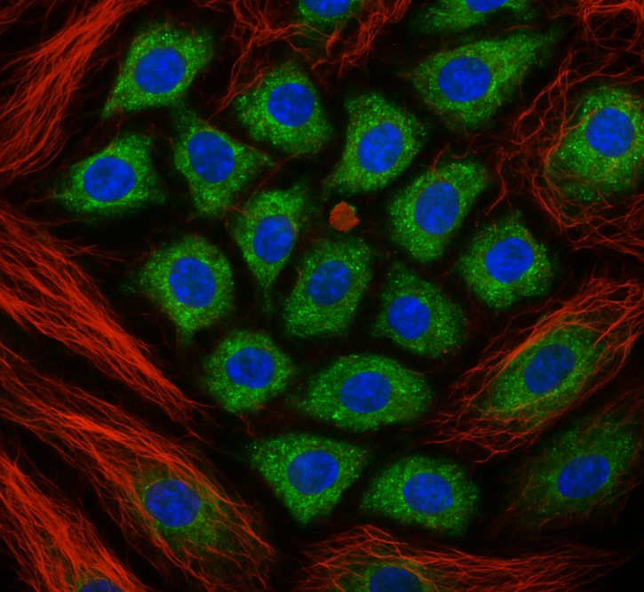
Seeing is believing, a well-known phrase holds excellent significance in science and in this context, optical microscopy is an important, widely accepted and non-invasive tool for direct visualization of microstructures of materials, biological specimens, and more. Confocal microscopy is a powerful optical imaging technique that facilitates the acquisition of high-resolution and high contrast images of samples when compared to conventional wide-field microscopy. This is achieved by employing a pinhole in front of the detector that spatially filters out-of-focus light from above and below the focal plane.
Confocal microscopy can provide better depth penetration and resolution in the axial direction through appropriate optical sectioning, thereby enhancing three-dimensional visualization, particularly useful for observing thick tissues like skin tissue, brain slices and plant cell walls, morphology and growth of spheroids and organoids, sub cellular localization of drug molecules and biological markers, and colocalization studies. It enables researchers to distinguish between autofluorescence and real fluorescence signals at the time of experiment directly, which is particularly helpful when imaging plant tissues, fungal or algae samples. Confocal microscopy allows live cell imaging, making it possible to observe dynamic processes such as cell division, migration, membrane dynamics and signal transduction.
Using FRET, FRAP, FCS, FLIM and FLIP, one can quantify the dynamics of interaction and diffusion coefficients of the components in the sample. The technique is helpful for examination of photophysical properties of synthesized dyes and nanomaterials like quantum dots, nanowires, and nanoparticles. With recent advancements, using super-resolution modalities, nanoscale organization and substructures of biomolecules can also be visualized.

FV4000
Enhance your images using the FLUOVIEW™ FV4000 confocal laser scanning microscope. Its cutting-edge imaging technology ensures greater image accuracy, providing researchers with more dependable data from their samples.
Applications & Specialities, Confocal, Microscopy, Products & Services, EVIDENT

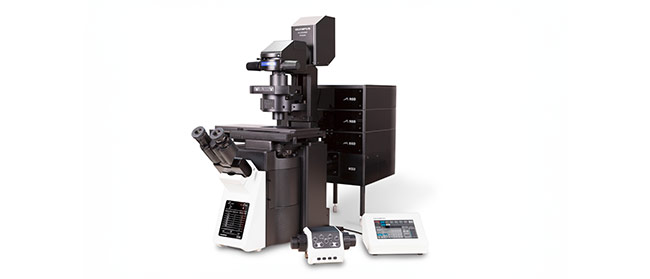
FVMPE-RS
With the Olympus FVMPE-RS, countless possibilities for deep tissue observation are finally realized. The system delivers unmatched high-speed imaging, essential for capturing the dynamic in vivo response, with fine laser control pinpointing the precise site for optimum excitation efficiency - even deep within the sample.
Applications & Specialities, Confocal, Instruments, Microscopes, Microscopy, Multiphoton Imaging, EVIDENT, Microscopy & Imaging
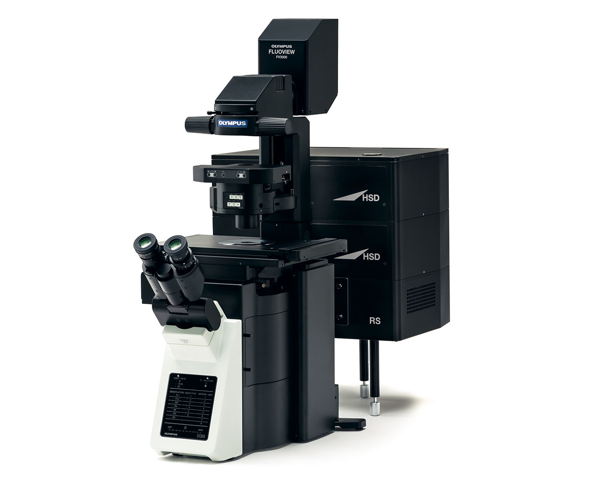
Olympus FV3000 Microscope
The FLUOVIEW FV3000 Series is designed to meet some of the most difficult challenges in modern science. With the high sensitivity and speed required for live cell and tissue imaging
Applications & Specialities, Confocal, Instruments, Microscopes, Microscopy, EVIDENT, Microscopy & Imaging
Testimonials & Reviews

Dr. Chhaya Chande, Professor & HOD, Microbiology
GGMCJJ Hospitals, Mumbai
“Ms. Megha Dhumal (Assistant Manager- Application) has done a satisfactory demonstration of the running of the Abbott Sample preparation machine model m2000sp and the Abbott RT-PCR machine model m2000rt. We appreciate the effort made by the DSS team under these difficult conditions to help our lab to carry out the imperative Covid-19 tests.”
Dr Sunil K Arora, Professor, Deptt of Immunopathology
PGIMER, Chandigarh
“We are using Confocal Microscope and one Fluorescence Microscope. Both are working fine. The after sales services by DSS have been excellent for functioning & upkeep of the microscopes. The applications support by experts from DSS is very useful. Keep it up!”
Dr Pramod Kumar Bajaj
MD, Spermprocessor Pvt Ltd
“Really excited to see the DSS Pathology solutions exhibition booth at APCON 2019 along with Magnus. We think all the upcoming technology had been displayed along with your efforts to make it Indigenous (Made in India) is highly appreciated. Wish you all the best. Keep it up!”
Dr. Sreejesh S, Associate Professor, Dept of Hematology
PGIMER, Chandigarh
“My experience with DSS so far has been very good till now. We are getting good support in both purchase as well as in troubleshooting. Very good experience with Mr Arun, Mr Manoj, Mr Mahesh and all others from the DSS team.”
Dr Sudha S Murthy, Department of Pathology and Laboratory Medicine
BIACH & RI, Hyderabad
“I am happy with DSS and associated with 19 years and use Dako antibody. Happy with Supply but need improvement.”
Dr S Radhika MD, PhD
Professor, Deptt. Of Cytology & Gynaec Pathology, PGIMER, Chandigarh
“PGI Cytology Dept. has had a long association with DSS- Olympus Microscopy Division. They have provided excellent services- after sales service. The product is also of very good quality. We have had no problems with their products and services are of very good quality.”
Dr Nuzhat Husain
RMLIMS, Lucknow
“Have been using Dako Reagants and Dako antibodies for a while. Services and products have been good and timely.”
Dr Minu Singh
Assistant Professor, PGIMER, Chandigarh
“MRC Holland MLPA products provided by DSS are of good quality, have never faced any quality issues with their product or shipping condition. They provide prompt response upon any query.”
Mr. Krishnani Professor, SGPGI, Lucknow
“My experience with DSS so far has been excellent for the last 30 years- sales and service experience. Microscope products are very useful and sturdy with high precision.”